Unveiling the Topography of the Eye: A Comprehensive Exploration of Tangential Map Corneal Topography
Related Articles: Unveiling the Topography of the Eye: A Comprehensive Exploration of Tangential Map Corneal Topography
Introduction
With enthusiasm, let’s navigate through the intriguing topic related to Unveiling the Topography of the Eye: A Comprehensive Exploration of Tangential Map Corneal Topography. Let’s weave interesting information and offer fresh perspectives to the readers.
Table of Content
- 1 Related Articles: Unveiling the Topography of the Eye: A Comprehensive Exploration of Tangential Map Corneal Topography
- 2 Introduction
- 3 Unveiling the Topography of the Eye: A Comprehensive Exploration of Tangential Map Corneal Topography
- 3.1 The Significance of Corneal Topography
- 3.2 Understanding Tangential Map Corneal Topography
- 3.3 Tangential Map Corneal Topography: A Closer Look
- 3.4 FAQs on Tangential Map Corneal Topography
- 3.5 Tips for Optimizing Tangential Map Corneal Topography
- 3.6 Conclusion
- 4 Closure
Unveiling the Topography of the Eye: A Comprehensive Exploration of Tangential Map Corneal Topography

The cornea, the transparent outer layer of the eye, plays a pivotal role in focusing light onto the retina. Its intricate shape, characterized by a subtle curvature, is crucial for optimal vision. Understanding the precise topography of the cornea is essential for diagnosing and managing various eye conditions, particularly refractive errors and corneal diseases. Tangential map corneal topography, a powerful diagnostic tool, provides a detailed visualization of the corneal surface, revealing its subtle variations and potential irregularities.
The Significance of Corneal Topography
Corneal topography is the process of mapping the surface of the cornea, generating a detailed representation of its shape and curvature. This information is invaluable in several ophthalmic applications, including:
- Refractive Error Assessment: Corneal topography helps determine the nature and severity of refractive errors like myopia, hyperopia, and astigmatism. This information guides the selection of appropriate corrective lenses or refractive surgery procedures.
- Corneal Disease Diagnosis and Monitoring: Corneal topography is instrumental in diagnosing and monitoring various corneal diseases, such as keratoconus, a progressive corneal thinning disorder, and corneal ectasia, a condition where the cornea bulges outward. Early detection and monitoring using corneal topography can facilitate timely intervention and prevent vision loss.
- Contact Lens Fitting: Corneal topography plays a crucial role in optimizing contact lens fitting. By providing a detailed map of the corneal surface, it helps practitioners select the appropriate lens size, shape, and material for optimal comfort and vision.
- Pre- and Post-Operative Assessment: Corneal topography is essential for pre- and post-operative evaluation in refractive surgery procedures, such as LASIK and PRK. It helps determine the suitability of the patient for surgery, monitor healing, and assess the effectiveness of the procedure.
Understanding Tangential Map Corneal Topography
Tangential map corneal topography is a specific type of corneal topography that utilizes a unique approach to visualize the corneal surface. Unlike other techniques that focus on the overall shape and curvature of the cornea, tangential mapping emphasizes the subtle variations in corneal curvature along the periphery of the cornea.
How it Works:
Tangential map corneal topography relies on a sophisticated instrument called a Placido disc topography system. This system projects a series of concentric rings onto the cornea. By analyzing the reflected patterns, the instrument calculates the corneal curvature at various points across the surface. The resulting data is then displayed as a tangential map, which highlights the subtle variations in corneal curvature along the periphery.
Advantages of Tangential Mapping:
- Enhanced Detection of Peripheral Irregularities: Tangential maps are particularly adept at detecting subtle irregularities in corneal curvature, especially in the peripheral regions. This is crucial for identifying early signs of corneal diseases like keratoconus, which often manifest initially in the periphery.
- Precise Astigmatism Assessment: Tangential maps provide a more precise assessment of corneal astigmatism, a condition where the cornea has an uneven curvature, leading to blurry vision. This information is vital for accurate prescription of corrective lenses or refractive surgery.
- Improved Contact Lens Fitting: Tangential maps offer valuable insights into the peripheral corneal shape, aiding in the selection of contact lenses that fit comfortably and provide optimal vision.
Tangential Map Corneal Topography: A Closer Look
Key Features:
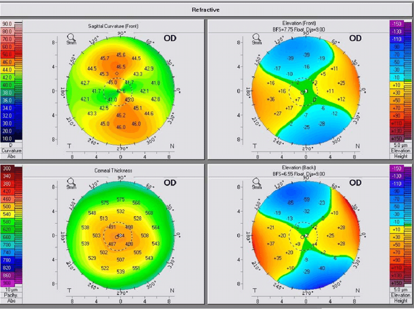
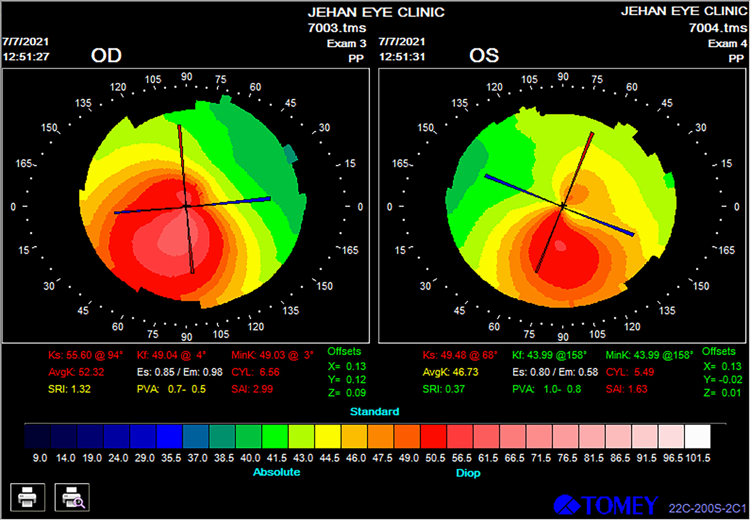
- Color-Coded Representation: Tangential maps typically use color coding to represent the different curvature values across the corneal surface. Red areas indicate steeper curvature, while blue areas indicate flatter curvature.
- Contour Lines: Contour lines, also known as iso-indices, connect points with similar corneal curvature. These lines provide a visual representation of the subtle variations in corneal shape.
- Peripheral Zone Emphasis: Tangential maps focus on the peripheral zone of the cornea, highlighting subtle changes in curvature that might be missed by other topographic techniques.
Interpretation of Tangential Maps:
- Regular Astigmatism: In regular astigmatism, the contour lines on a tangential map are typically symmetrical and parallel, indicating a predictable pattern of corneal curvature.
- Irregular Astigmatism: Irregular astigmatism, often associated with corneal diseases, results in asymmetrical and distorted contour lines on a tangential map.
- Keratoconus: Tangential maps in keratoconus patients often exhibit characteristic patterns, such as a steepening of the central cornea and a flattening of the periphery, leading to a "cone-shaped" appearance.
FAQs on Tangential Map Corneal Topography
Q: Is tangential map corneal topography a painful procedure?
A: No, tangential map corneal topography is a non-invasive procedure that does not involve any discomfort. It is similar to other corneal topography techniques, where a series of light patterns are projected onto the cornea, and the reflected patterns are analyzed.
Q: How often should I have tangential map corneal topography performed?
A: The frequency of tangential map corneal topography depends on individual factors, including the presence of any corneal diseases or risk factors. Your ophthalmologist will determine the appropriate frequency based on your specific needs.
Q: What are the limitations of tangential map corneal topography?
A: While tangential map corneal topography is a powerful tool, it does have some limitations. The accuracy of the measurements can be affected by factors such as eye movement, tear film stability, and the presence of corneal opacities.
Q: Can tangential map corneal topography be used for all eye conditions?
A: Tangential map corneal topography is particularly useful for assessing refractive errors and corneal diseases. However, it may not be suitable for all eye conditions, such as cataracts or retinal diseases.
Q: What are the alternatives to tangential map corneal topography?
A: Other corneal topography techniques include Placido disc topography, Scheimpflug imaging, and corneal elevation mapping. The choice of technique depends on the specific clinical application and the information required.
Tips for Optimizing Tangential Map Corneal Topography
- Eye Relaxation: It is important to relax your eyes during the procedure to minimize eye movement, which can affect the accuracy of the measurements.
- Tear Film Stability: Ensure that your eyes are well-lubricated with artificial tears if necessary to maintain tear film stability.
- Proper Eye Alignment: Ensure that your eyes are properly aligned with the instrument to obtain accurate measurements.
- Collaboration with Your Ophthalmologist: Discuss your concerns and expectations with your ophthalmologist to ensure the appropriate use of tangential map corneal topography.
Conclusion
Tangential map corneal topography is a valuable diagnostic tool that provides detailed information about the corneal surface, particularly the subtle variations in curvature along the periphery. This information is crucial for accurate diagnosis and management of various eye conditions, including refractive errors and corneal diseases. By providing a comprehensive understanding of the corneal topography, tangential mapping empowers ophthalmologists to make informed decisions regarding treatment options and optimize patient outcomes. As technology continues to advance, tangential map corneal topography will undoubtedly play an increasingly significant role in improving eye care and preserving vision.


Closure
Thus, we hope this article has provided valuable insights into Unveiling the Topography of the Eye: A Comprehensive Exploration of Tangential Map Corneal Topography. We hope you find this article informative and beneficial. See you in our next article!